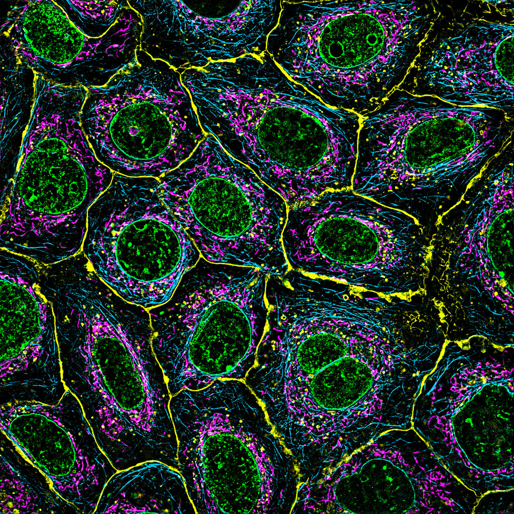

The Zeiss Lattice SIM 5 fluorescence microscope forms part of Australian Institute for Microbiology and Infection (AIMI) contribution to super-resolution microscopy in the MIF. The system is an inverted super-resolution structured illumination microscope suitable for live and fixed samples. The Lattice SIM technology in combination with SIM2 image reconstruction algorithms can achieve resolution down to 60 nanometres. SIM apotome mode is also available for fast optical sectioning of large samples for high spatial resolution. A stage top incubator with both temperature and CO2 control allows for live cell imaging.
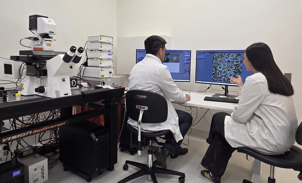
Microscope
- Axio Observer inverted microscope with motorised objective turret and filter turret
- Lattice SIM 5 super-resolution illumination head
- AI Sample Finder for sample navigation
- Hardware (Definite Focus) and software based focusing methods

Objectives
| Objective | NA | Technique | Immersion | WD (mm) |
| EC Plan-Neofluar 10x | 0.3 | SIM Apotome | Air | 5.2 |
| Plan-Apochromat 20x | 0.8 | SIM Apotome | Air | 0.55 |
| Plan-Apochromat 40x | 1.4 | SIM Apotome | Oil | 0.13 |
| Plan-Apochromat 63x | 1.4 | Lattice SIM | Oil | 0.19 |
| Plan-Apochromat W Korr 63x | 1.2 | Lattice SIM | Water | 0.28 |
- Auto-immersion unit for water objective
Light sources
- Transmitted light and epi-fluorescence (for observation)
- Laser lines include 405nm, 488nm, 561nm, 640nm
Filters
SR HE LBF 405/488/561/642
| Ex | BP | Em |
| 200-405 | 405 | 419-470 |
| 200-488 | 488 | 503-546 |
| 200-561 | 561 | 576-617 |
| 200-642 | 642 | 657-800 |
SR HE BF 415-475 + BP 500-55
| Ex | BP | Em |
| 200-405 | 405 | 419-470 |
| 200-488 | 488 | 503-546 |
| 200-642 | 642 | 657-800 |
SR HE BF 500-575 + BP 575
| Ex | BP | Em |
| 200-488 | 488 | 503-546 |
| 200-561 | 561 | 576-617 |
SR HE BF 415-475 + LP 655
| Ex | BP | Em |
| 200-405 | 405 | 419-470 |
| 200-642 | 642 | 657-800 |
Detectors
- Hamamatsu Fusion BT 5.3 Megapixel sCMOS camera
Software and Processing
Zen v3.14 including:
- 2D Burst mode using a rolling window approach for live cell imaging up to 255 fps.
- 3D Leap mode for digital sectioning to reduce imaging time and light exposure.
A dedicated processing computer is available for on-the-fly image reconstruction using SIM² image reconstruction algorithms. A digital SIM point spread function is first constructed and subsequentially used in iterative deconvolution. This SIM² algorithm is superior to conventional one-step image reconstruction methods in terms of resolution, sectioning, and robustness.