Lecturer:
Catherine Gorrie Faculty:
Science Project:
Virtual Microscopy for teaching Histology and the biomedical sciences Keywords:
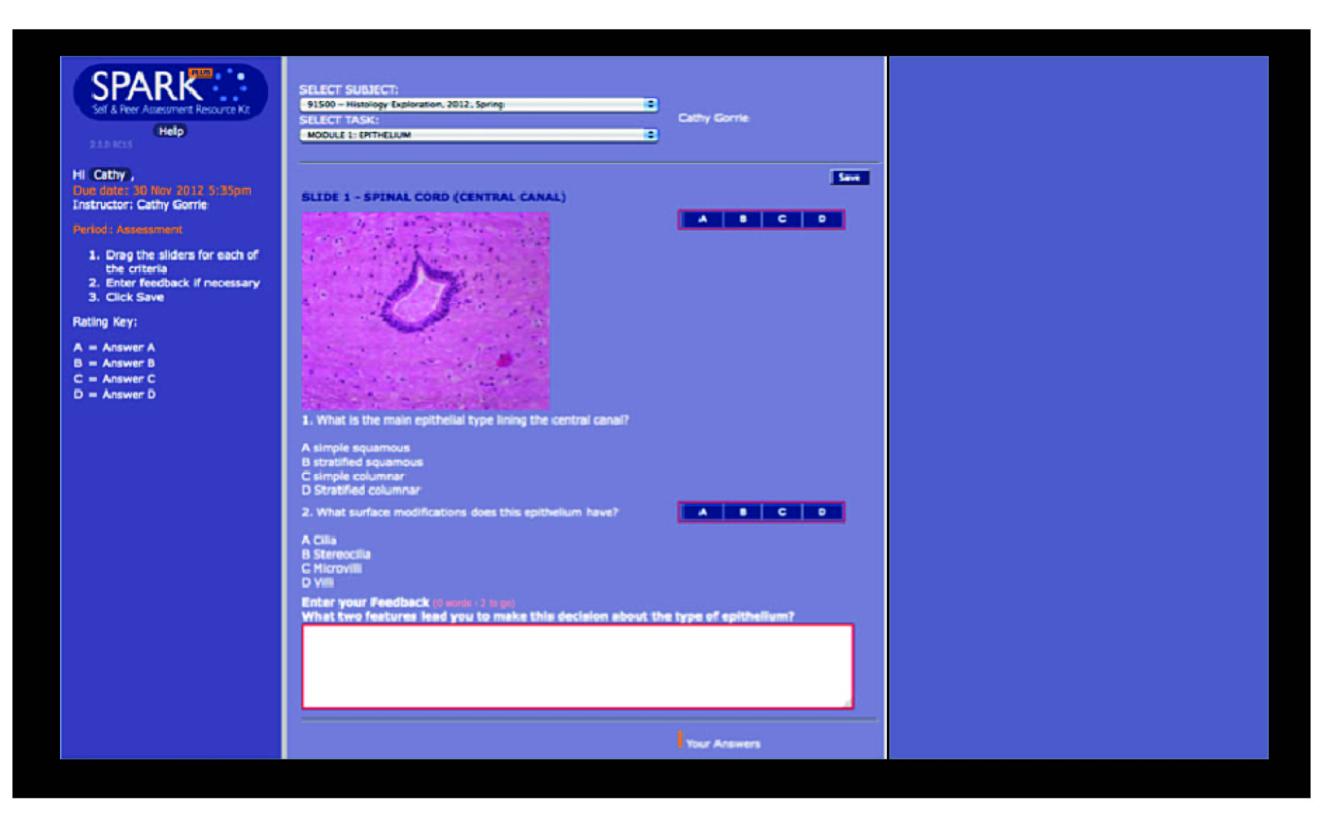
Histology, microscope simulation, SPARKplus, self-directed learning activities, revision | | Using a virtual microscope to increase students’ access to and identification of microscope slides, Dr Catherine Gorrie’s project is extending conventional microscopy learning in Science courses. What the project is aboutDr Gorrie is developing an online ‘virtual microscope’ to provide students with a resource tool for use prior to and after laboratory classes and lectures in Histology. Students will be able to, for example, compare and review the virtual microscope slide images with those viewed looking down the actual microscope in their laboratory class.Why the project is being introducedStudents’ access to microscopes is limited outside laboratory class. This project will therefore support and enhance current curriculum by providing students with a ‘virtual microscope’ they can access from home to identify, revise and develop understanding of histology-related ideas. It will also provide greater opportunities for students to work together, discuss and test out their ideas. How the project is being implementedThe ‘virtual microscope’ is being implemented through the SPARKplus system. Dr Gorrie has developed each module on SPARKplus to cover one topic and take 10-20 minutes to complete. Students are provided with online assistance via a brief introductory tutorial explaining the SPARKplus system, how to access the images and complete the modules. Students use the system to provide and receive immediate feedback through images, questions and comments.
Based on her students’ completion of the online modules, Dr Gorrie is able to review which parts of the histology slides they understood or didn’t quite grasp. She then targets the trouble area(s) in that week’s lecture and practical class, instead of repeating already understood concepts or other content.
Having access to slides is difficult in Histology, with only one student at a time being able to look through the microscope in class. Dr Gorrie’s virtual microscope changes this by allowing any student to instantly view any of those slides from home. Based on their familiarity with those ‘digital slides’, students can now also work in groups to easily discuss what is being looked at under the microscope in their laboratory classes. One student noted the benefits of, the ‘virtual microscope’ in this way: it “helped me in preparing what to look for in the next day’s lab class as I’m always unsure of what to look for during pracs”. Challenges and considerationsInitially, the quality of the images under the ‘virtual microscope’
needed improving to increase clarity for analysis and identification.
This problem was solved, and now Dr Gorrie is looking at having a zoom function developed.
Dr Gorrie recommends not only spending time on presenting new and novel concepts to the students but also considering how to use other resources to guide that conceptual development and increase their participation:
“I think I expected students to embrace this from the start and some did, but I think many were just bewildered, especially as it was not
assessed. I have a better idea of how the software works now, and
will be able to develop and integrate everything for students better
next year.”
Exploring further
SPARKplus
University of Michigan. Medical Histology and Virtual Microscopy
UTS Library. A brief introduction to Open Educational Resources.
Delving even deeper
JISC. A Guide to Open Educational Resources
Learning2014 on Pinterest. Discipline-specific examples of Open Educational Resources
|