



The Nikon A1R is an inverted confocal microscope with two standard PMT detectors and two standard GAaSP detectors. A separate 32 channel spectral detector, and transmitted light PMT detector are also available. The system is inside an environmental chamber with temperature, CO2 and humidity control.
Microscope
- Inverted TiE Stand
- Motorized x-y stage
- Piezo z stage
- Perfect Focusing System
Nikon objectives
The MIF has a range of Nikon objectives that can be installed on Nikon microscopes. Please contact the MIF team if you need a specific objective installed on a microscope prior to your imaging session.
| Objectives | NA | Tech | WD/mm | Imm. | PFS | Material Ref. |
|---|---|---|---|---|---|---|
| 4X Plan Apo 𝝺 | 0.2 | BF | 20 | Air | Y | MRD00045 |
| 4X Plan Fluor DL F | 0.13 | PhL | 16.4 | Air | N | MRH20041 |
| 10X Plan Fluor DLL | 0.3 | Ph1 | 16 | Air | Y | MRH10101 |
| 10X Plan Apochromat 𝝺 | 0.45 | DIC | 4 | Air | Y | MRD00105 |
| 20x Plan Apo lambda DM | 0.75 | Ph2 | 1 | Air | Y | MRD00205 |
| 20X Apo 𝝺S | 0.95 | 0.95 | Water | Y | MRD77200 | |
| 20X Plan Fluor DM ELWD | 0.45 | Ph1 | 8.20 - 6.90 | Air | Y | MRH08230 |
| 20X S Plan Fluor LWD | 0.7 | 2.3 - 1.3 | Air | Y | MRH08250 | |
| 40X Plan Apochromat 𝝺DM | 0.95 | Ph2 | 0.21 | Air | Y | MRD30405 |
| 40X S Plan Fluor ADM ELWD | 0.6 | Ph2 | 3.60 - 2.80 | Air | Y | MRH48230 |
| 40X Apo 𝝺S chromatic correction:405-850nm | 1.25 | 0.18 | Water | Y | MRD77400 | |
| 40X Plan Fluor H N2 | 1.3 | DIC | .24 | Oil | Y | MRH01401 |
| 60X APlan Apochromat VC | 1.2 | 0.29 | Water | Y | MRD07602 | |
| 60X Plan Apochromat 𝝺DM | 1.4 | DIC | 0.13 | Oil | Y | MRD31605 |
| 100X Plan Apochromat 𝝺 | 1.45 | 0.13 | Oil | Y | MRD01905 | |
| 100X Plan Apo 𝝺DM | 1.45 | Ph3 | 0.13 | Oil | Y | MRD31905 |
Lasers
- 405, 457, 514, 488, 561, 640 nm
Scanners
- Galvano scanner up to 24 Hz
- Resonant scanner up to 420 Hz (512 x 512)
Software
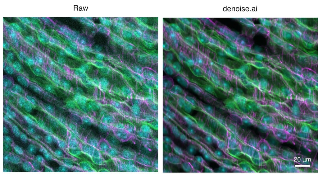
NIS Elements including FRET module, JOBS for automated acquisition and real time analysis workflows, NIS.ai denoise and deconvolution algorithms for image processing.
*Mother of Pearl
Maximum intensity projection of whole placental organoid immunolabelled for E-Cadherin (orange), Human Leukocyte Antigen-G (chartreuse), Syndecan-1 (light blue), and counterstained with DAPI (pink). Placental organoids were retrieved from their matrix and grown in suspension for 12 days. Whole organoids were fixed, blocked, immunolabelled, and optically cleared. Whole organoids were mounted on slides and imaged to a depth of 60um using a Nikon A1R Confocal microscope using a 20X NA0.45 objective with 0.8um z-steps. Image was denoised using Denoise.Ai.