


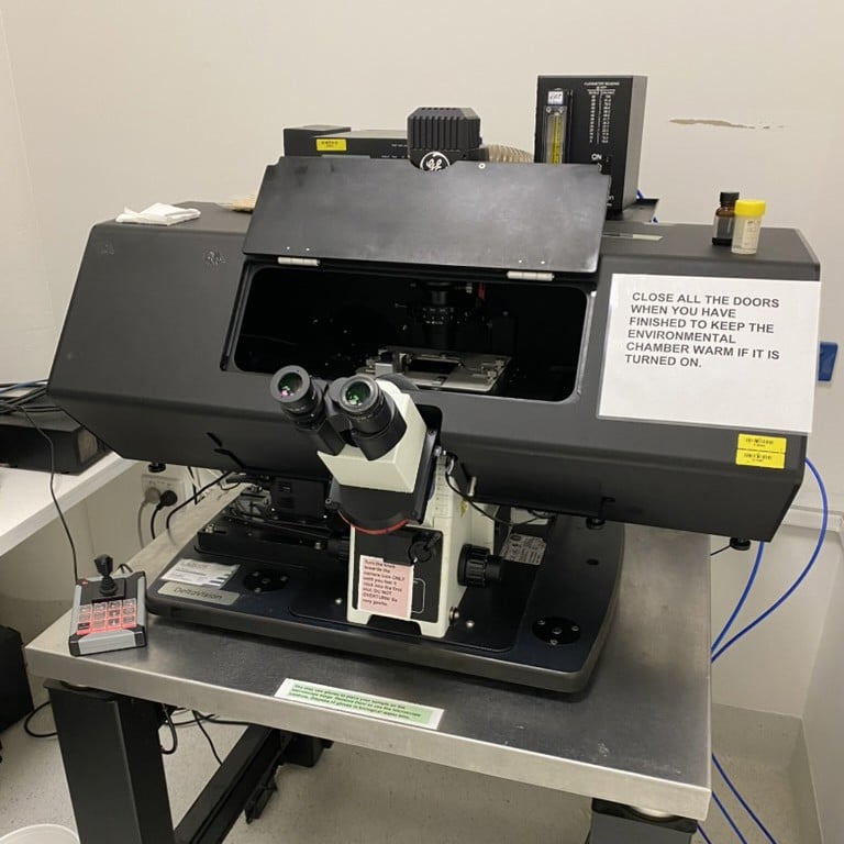
DeltaVision Elite with Phase Contrast optics
Inverted Widefield fluorescence deconvolution microscope.
- Environmental control chamber with CO2 and temperature control.
Lenses
| Objective | NA | Tech | WD/mm | Imm. | PFS | Material Ref. |
|---|---|---|---|---|---|---|
| 10X U-Plan S-Apo | 0.4 | 3.1 | Air | Y | 1-U2B823 | |
| 20X U-Plan FL LWD | 0.45 | Ph1 | 6.6 | Air | Y | 1-U2B375 |
| 40X UApo | 1.30 | 0.1 | Oil | Y | 1-UB768 | |
| 60X Plan Apo N | 1.42 | DIC | 0.15 | Oil | U | 1-U2B933 |
| 60X LCPLPL | 0.7 | Ph2 | 0.15 | Air | Y | 1-UC351 |
| 100X Plan Apo | 1.4 | Ph3 | 0.13 | Oil | Y | 1-U2B836 |
Filters
There are three polychroics available. Within each polychroic, the four excitation and emission filters can be independently chosen.
D-T-F-C
| Filter | Ex/nm | Em/nm |
|---|---|---|
| DAPI | 381-412 | 420-456 |
| FITC | 464-492 | 500-523 |
| TRITC | 531-565 | 573-611 |
| Cy5 | 619-646 | 654-700 |
D-F-mCh-C
| Filter | Ex/nm | Em/nm |
|---|---|---|
| DAPI | 381-412 | 420-456 |
| FITC | 464-492 | 500-549 |
| mCherry | 557-590 | 598-617 |
| Cy5 | 625-646 | 654-700 |
C-Y-mCh
| Filter | Ex/Nm | Em/nm |
|---|---|---|
| CFP | 400-453 | 463-487 |
| YFP | 497-527 | 537-549 |
| mCherry | 557-592 | 602-662 |
*Fluorescent visualisation of Staphylococcus aureus SH1000-GFP microcolony formation
This fluorescence micrograph captures the early growth behavior of GFP-tagged Staphylococcus aureus SH1000 after 6 hours of incubation. Planktonic cells were removed prior to imaging to visualize only adherent bacterial cells. Distinct fluorescent clusters reveal active cell proliferation and the onset of microcolony formation, highlighting the transition from planktonic cells to structured surface-associated communities.