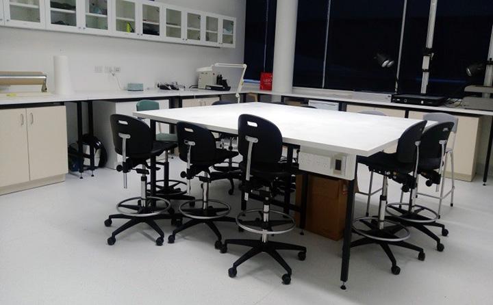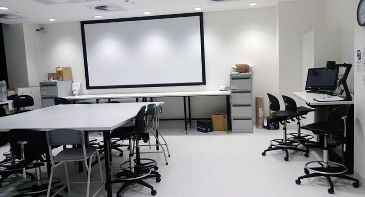

The Forensic suite has various specialised instruments available for forensic applications such as fibres analysis, finger marks detection, chemical trace analysis (including paint, fibres, glass, fire and explosive residues), document examination, drug analysis and microscopy analysis. Contact Dr Linda Xiao for more information about the instruments and facilities.
List of the major instruments in the Forensic suite:
Fourier Transform Infrared (FT-IR) Spectroscopy
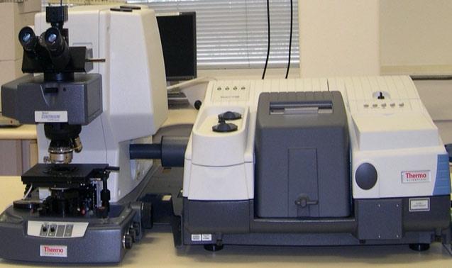
The Thermo Scientific Nicolet 6700 FTIR spectrometer is connected to a Nic-Plan IR microscope for either transmission or reflections microscopy. Infrared absorption by carbon dioxide and water vapour in ambient air can be minimised by purging the optics with dry nitrogen.
Applications of FTIR spectroscopy include helping to identify unknown materials, determine the quality of a sample and quantify components in a mixture. The instrument has a spectral range of 7,400 - 375 cm-1 using KBr beam splitter and 0.9 cm-1 standard optical resolution.
Raman Spectroscopy
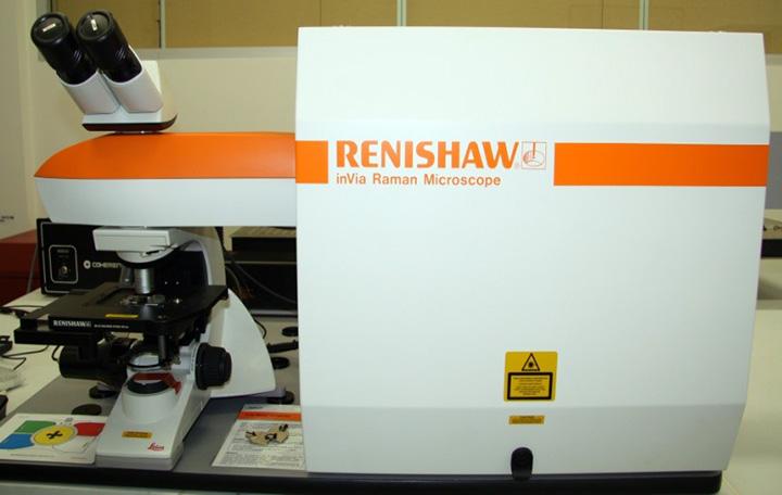
A Renishaw inVia Raman spectrometer system (Gloucestershire, UK) equipped with a Leica DMLB microscope (Wetzlar, Germany) and a 17mW at 633 nm renishaw helium neon laser source.
The application of Raman spectroscopy includes inorganic, polymer, pharmaceutical , semiconductors, art, archaeology and biotech areas analysis, especially in forensic and process analysis.
Infrared Chemical Imaging
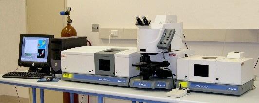
Infrared chemical imaging is carried out using a Digilab Stingray system consisting of an FTS7000 infrared spectrometer, a UMA600 infrared microscope, a Lancer 64x64 MCT focal plane array detector, and the Stingray large sample (imaging) accessory.
The instrument is also equipped with a single point MCT detector (microscope), a DTGS detector (bench), an MTEC photoacoustic detector, and hot and cold stage capabilities (-200 to +1500 degC).
Microspectroscopy
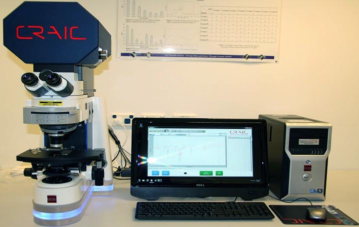
The 20/20 PV™ Craic Microspectrometer can take spectra and images of microscopic samples from the deep ultraviolet to near infrared (200-2100nm) with one seamless operation. It can acquire microspectra™ and images in absorbance, reflectance, and fluorescence modes.
The applications of Microspectrometers include identification and quantification of microscopic samples ranging from the microfluidic kinetics, matching fibers or paints, the qualification of gems or coal, the determination of the color of ink or paint or even the analysis of great works of art. As such, the microspectrometer is a highly flexible instrument with many different applications.
Video Spectral Comparator
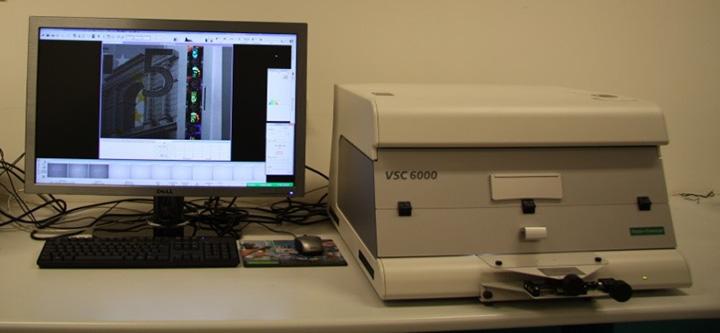
The VSC6000/HR (Foster+Freeman) is a comprehensive digital imaging system with an extensive range of features and applications. Techniques include examination in the visible and infrared regions of the spectrum carried out with incident and transmitted UV, visible and infrared illumination up to 1000 nm.
Equipped with a high resolution digital colour video camera the VSC offers 5M pixel digital Firewire camera 2584 x 1956 viewable pixels with x22 motorised zoom output providing excellent image quality. Software features include casework management; image integration, processing and measurement; colour measurement and archiving facilities.
Polilight and Poliview x2
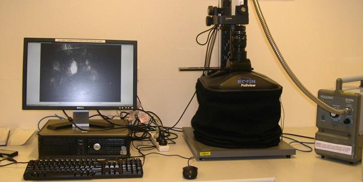
The Polilight (Rofin Australian) is the leading forensic light source for both scene and laboratory applications. The instrument produces 14 light bands in the UV, visible and infrared range delivered via a flexible light guide. Applications include latent fingerprint and document examination, as well as for trace evidence such as fibers, bloodstains, hairs, paint chips, semen, saliva and some illicit drugs.
The Poliview (Rofin Australia) is an integrated image detection and enhancement system that incorporates the Polilight. The system includes a CCD camera with 1024 x 1024 pixel resolution and a 14x zoom lens, capable of 10 minute integration times and employs all available optical techniques - fluorescence, reflection, absorption, and transmission imaging.
Ballistics Comparison Microscope
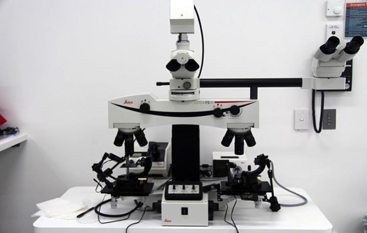
The Leica DMC comparison microscope is ideally equipped for forensic examination; allowing similarities and differences between two bullets or cartridges to be compared. Split-image and superimposed image comparisons are possible, with the microscope being equipped with a CCD camera for photography and video documentation.
A key feature of the microscope is that the light is focused through the objective, dramatically increasing definition of surface features visible to the eye. The end magnification ranges from 4x to 80x, corresponding to object field sizes of 50mm to 2.5mm. A large variability of clamping and mounts for different objects are available.
Fibre Comparison Microscope
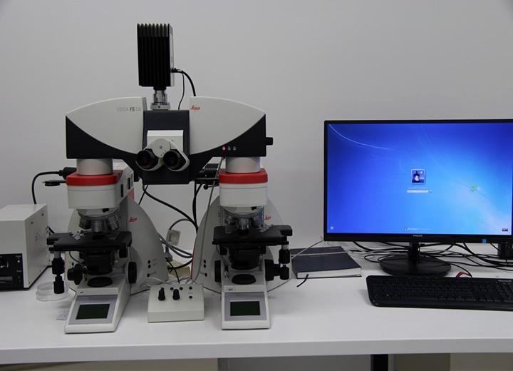
Leica Fibre comparison Microscope enables trace evidence comparisons in forensic science laboratories. Comparison bridge integrates tilting binocular tube that promotes ease of reach with natural body position. Automation and motorization of comparison bridge allow one-button activation of comparison methods and movement of image splitting line.
Glass Refractive Index Measurement
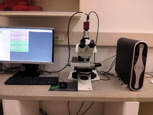
GRIM 3 (Foster+Freeman) is a machine used to compare glass fragments to see if they match. The fragments of glass are crushed and placed on a glass microscope slide. A few drops of a special type of silicon oil is placed on the crushed glass fragments and then the slide is placed in a heated chamber, known as a hot stage which is placed under a microscope. This resulting image from the microscope is fed via a camera attachment, to a computer. The silicon oil on the slide is heated up slowly by the hot stage which alters the refractive index of the oil. When its refractive index is equal to that of the glass, the glass fragments become invisible! The computer can determine better then the human eye at which point the glass becomes invisible and records the optimum match temperature. The refractive index is then calculated from two match temperatures made by the computer.
Crime Lite x3
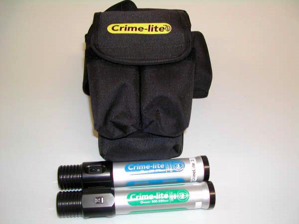
Using advanced LED technology, Foster Freeman have created a range of compact, battery powered, high-intensity forensic light sources for locating evidence at the scene of a crime.
Crime-lites provide narrow bandwidth illumination in the following wavelengths:
- Blue Crime-lite® with a wavelength peak of 450nm (10% bandwidth 430-470nm) for general search for body fluids.
- Green Crime-lite® with a wavelength peak of 520nm (10% bandwidth 500-550nm) for DFO treated fingerprints.
Humidity Cabinet x2
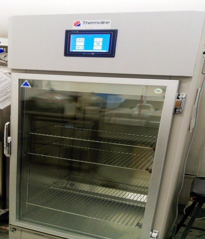
The Thermoline Humiditherm temperature and humidity chamber has been designed to provide control of temperature between +10°C and 60°C with a humidity range of 30 to 90% RH.
Forensic Cyanoacrylate Cabinet x2
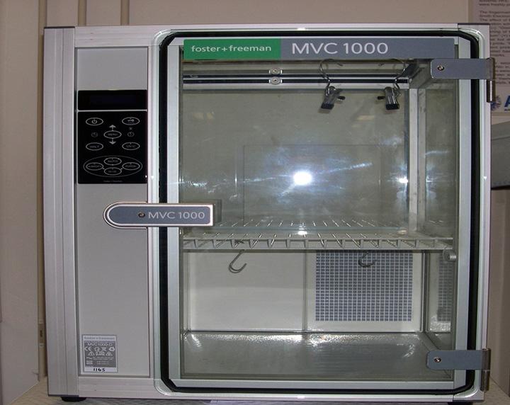
Safe Automated Development of Latent Fingerprints for High Quality Evidence
The FCC171 Forensic Cyanoacrylate Cabinet (Foster+Freeman) was developed by experienced forensic scientists to enable the safe and efficient development of latent fingerprints on non-porous surfaces using cyanoacrylate vapour at atmospheric pressure.
Vacuum Metal Deposition (VMD)
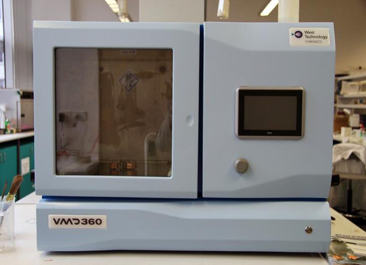
VMD 360 (West Technology) is a powerful forensic technique that uses the sequential vacuum deposition of gold and zinc to develop latent fingerprints. VMD can be used on a wide range of non-porous and semiporous exhibits including flexible plastic packaging, plastic bottles, glass, fabrics, firearms and glossy paper or magazines. The technique is quick (typically less than 15 minutes) and produces high quality images with ‘3rd’ level detail of pores and ridge shapes. The developed prints can be photographed immediately.
Electrostatic Imaging System for Detecting Indented Writing Analysis (ESDA 2)
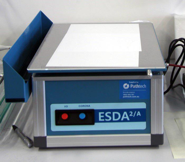
ESDA 2 (Foster + Freeman) works by creating an electrostatic image of indented writing, which is then visualised by the application of charge sensitive toners. The sensitive imaging process reacts to sites of microscopic damages to fibres at the surface of a document, which have been created by abrasive interaction with overlying surfaces during the act of handwriting.
UV-VIS Spectroscopy
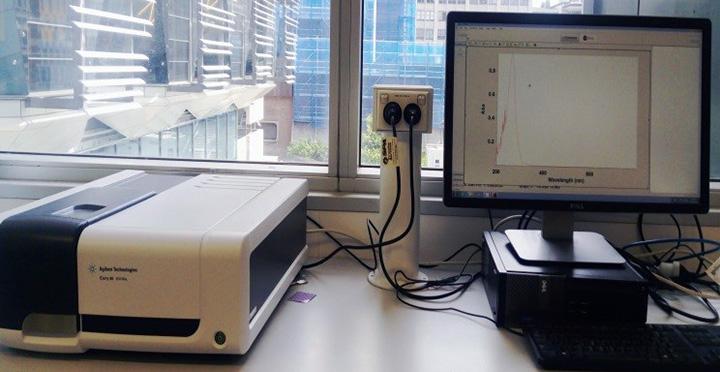
The Agilent Cary 60 UV-Vis spectrophotometer comprises a double beam, Czerny-Turner monochromator, 190–1100 nm wavelength range, 1.5 nm fixed spectral bandwidth, full spectrum Xenon pulse lamp single source with exceptionally long life, dual silicon diode detectors, quartz overcoated optics, scan rates up to 24,000 nm/min, 80 data points/sec maximum measurement rate, nonmeasurement phase stepping wavelength drive, room light immunity, central control by PC with Microsoft® Windows® operating system. Supported by GLP software.
Crime Scene Simulation Lab
Forensic students at UTS Science have access to a unique Crime Scene Simulation Lab. Set up like a city apartment, this new facility provides the students with an interactive and more realistic view in analysing the modern crime scene.
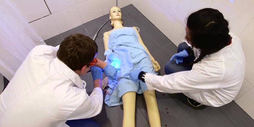
Forensic students at UTS Science have access to a unique Crime Scene Simulation Lab. Set up like a city apartment, the lab provides students with practical experience in crime scene investigations.
[Instrumental music]
[Close up of someone in lab coat scanning pass and opening door, then close up of sign that says Crime Scene Simulation Lab.]
[Forensic student in mock dining room dusts for fingerprints; student in mock lounge room takes a photo of evidence.]
Custom designed virtual city apartment
[Three forensic students sit at bench with three computers examining photos of evidence and discussing findings.]
Training aspiring forensic scientists and crime scene investigators
[Close up of students discussing photos.]
Simulating real-life crimes
[A forensics student measures the length of a suspect’s shoe; close up of a suspect’s shoe with a tape measure next to it]
Break and enter
[The forensic student takes a photo suspect’s shoe in mock break enter crime scene.]
Suspicious circumstance
[Two forensic students in a mock bedroom remove covers from bed where a dummy body lays.]
Everyday settings where crimes typically occur
[Two forensic students in mock crime scene in dining room dust a glass for finger prints and use a blue light to examine evidence.]
Teaching fingerprint detection
[Close up of student dusting dining room table for finger prints.]
[Student rolls out yellow crime scene tape over mock bathroom crime scene while another student uses a blue light to look for evidence.]
Detecting blood hidden in crime scenes
[Second student joins one in bathroom as blue light shows mock blood in crime scene.]
[Close up to student in bedroom scene collecting evidence]
[Two students in mock study crime scene with dummy body look for evidence]
Working collaboratively
[Close up of waste bin where student finds gun and places into evidence bag.]
[Second student uses magnifying glass and takes notes.]
Developing new detection methods
[Kitchen shows mock drug lab where two students take photos and collecting evidence]
New human identification techniques
[Close up of student taking a swab of substance in glass container]
World leading forensics education
[Close up of student in mock bedroom looking at brochures and cards left on floor]
You can explore the Crime Scene Simulation spaces in the below link that was generated by 360 degree imaging capabilities that we have here at UTS.
Explore the Crime Scene Simulation Lab
Search Lab x2