


- Posted on 25 Aug 2026
- 6-minute read
A new artificial intelligence platform uses images from the back of the eye to simplify the diagnosis of critical diseases and injuries.
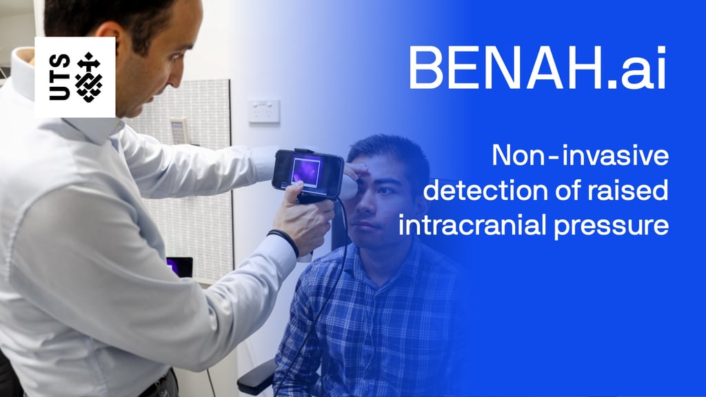
BENAH.ai combines precision retinal imaging with advanced artificial intelligence to aid identification of abnormal pressure on the brain faster and less invasively than existing diagnostic tools.
In 2009, a 12-year-old boy fell off his bike and hit his head on the pavement in Maryborough in central Victoria. After experiencing headaches, his mother took him to the local hospital where he was diagnosed with pressure on the brain.
The doctor on duty had to act quickly, using a drill from the maintenance room to relieve the pressure and bleeding on the boy’s brain with guidance over the phone from a Melbourne neurosurgeon.
Thanks to the quick help, the boy could then be airlifted to a major children’s hospital and was safely released in time for his 13th birthday.
“That story really sat with me,” said Associate Professor Mojtaba Golzan, an expert in vision science from the UTS School of Clinical and Health Sciences.
"If doctors had been able to detect the pressure on his brain earlier, they would have been able to make arrangements to transfer him to a larger hospital."
It’s an experience Golzan has taken into developing a new technology BENAH.ai that combines retinal imaging with artificial intelligence to identify intercranial pressure quickly and enable triaging for more detailed diagnosis.
“When somebody presents to an emergency department with a headache, clinicians need to quickly rule out whether they've got dangerous pressure building up within the brain,” he said.
“Currently, it can take a couple of hours or a couple of days to rule that out using brain imaging and lumbar punctures. With our technology, we’re trying to get that result in a couple of minutes.”
BENAH.ai is a software-as-a-medical-device that takes images from the back of the eye and puts them through an artificial intelligence platform which analyses them against features linked to abnormal brain pressure.
It then provides a probability score of how likely it is the patient has abnormal brain pressure.
“Swelling of the optic disc or optic nerve head – the medical term for it is papilledema – is associated with an increase in intracranial pressure,” Golzan said.
“But identifying it is not as simple as scanning a patient’s eye. Not all swelling of the optic nerve is caused by intracranial pressure, and not all pressure leads to a swelling in the eye.”
“It’s very difficult even for medical specialists and many emergency clinicians can struggle to fully understand the complexities of identifying it."
"They often need to bring in ophthalmologists or neurologists, which takes time.”
“That’s where our technology comes in – inspired by the Persian word BENAH (“bee.naa”) meaning “to see”.”
The artificial intelligence driving BENAH.ai is being developed to identify the key visual features in the back of the eye that correlate to increased brain pressure.
They have drawn on a large dataset of eye imaging and diagnostic data from historical patients from across various clinical sites to build and refine their models.
We found 5 distinct visual features in the eye correlated with pressure. Based on a panel of these, we could determine the likelihood of a patient having abnormal brain pressure.
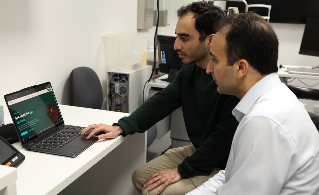
The portable eye scanning and imaging tool for the system, initially prototyped with help from UTS Rapido, is being developed in partnership with a hardware company, oDocs Eye Care.
The hardware has been approved for use by the Therapeutic Goods Administration.
Side-by-side with the hardware, the software for the benah.ai is honed to improve its accuracy ahead of it being trialled in emergency departments.
The AI model has demonstrated a sensitivity of 91 per cent in patients with confirmed intercranial pressure, meaning it correctly identified the condition in 9 out of 10 cases that have been fully diagnosed.
The model is currently being validated in a proof-of-concept clinical trial across two major Sydney hospitals with the aim of completing 100 patient assessments.
The team is continuing to refine the model as the clinical trial progresses, with the goal of further reducing missed cases.
“Diagnosing papilledema and its causes is incredibly difficult. Very few emergency clinicians are confident in using the instruments used to look at the back of the eye. Even neuroimaging like CT scans and MRIs can miss raised intercranial pressure,” Golzan said.
The next step after optimising the AI platform will be a dedicated trial in emergency departments to assess how the technology integrates into frontline clinical workflows ahead of regulatory submission.
“We need to show its going to expedite their processes, save their time and save patients from getting extra radiation or a needle into their spine,” said Golzan.
“If we can save just one patient from going through an invasive procedure, I’ll have achieved my mission. It's all about the patients."
"The driver for me is being able to give people alternative options where they can get quick and easy detection on the spot.”
Find out more
Transforming images and video into diagnostic insights for retinal diseases with confidence and speed.
Researcher
Deputy Head Of School, Research, Faculty of Health
Artificial intelligence ushers in new era of personalised medicine
Biomedical startup 23Strands is working with UTS Professor Jie Lu to use artificial intelligence to revolutionise personalised medicine.

The heart unbreakers
How bioprinted heart patches can revolutionise cardiac care.
Printing a cure for pre-eclampsia
Could 3D-printed mini placentas offer more effective treatments for pre-eclampsia? Researchers are investigating better, safer ways to study the placenta and...
Taking smarter, faster vaccines from lab to production
UTS is creating a pipeline of homegrown next-generation vaccines for Australia.
Artificial intelligence ushers in new era of personalised medicine
Biomedical startup 23Strands is working with UTS Professor Jie Lu to use artificial intelligence to revolutionise personalised medicine.
The heart unbreakers
How bioprinted heart patches can revolutionise cardiac care.
Printing a cure for pre-eclampsia
Could 3D-printed mini placentas offer more effective treatments for pre-eclampsia? Researchers are investigating better, safer ways to study the placenta and...
Taking smarter, faster vaccines from lab to production
UTS is creating a pipeline of homegrown next-generation vaccines for Australia.